 |
|
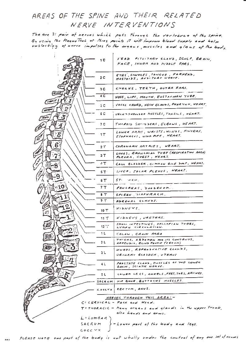
The
spinal
cord is about the size of a mans' little finger, the image
above is a greatly enlarged representation with needle to
scale overlayed. The outer layers of the spinal
cord are called the Meninges and are microscopically thin,
the needle is passed through these layers in to the space
which contains the cauda equina nerves and this is where
they injected Myodil.
|
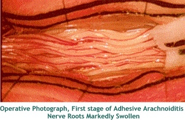
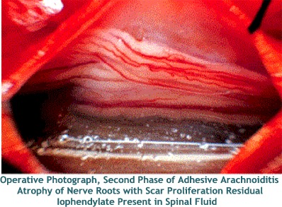
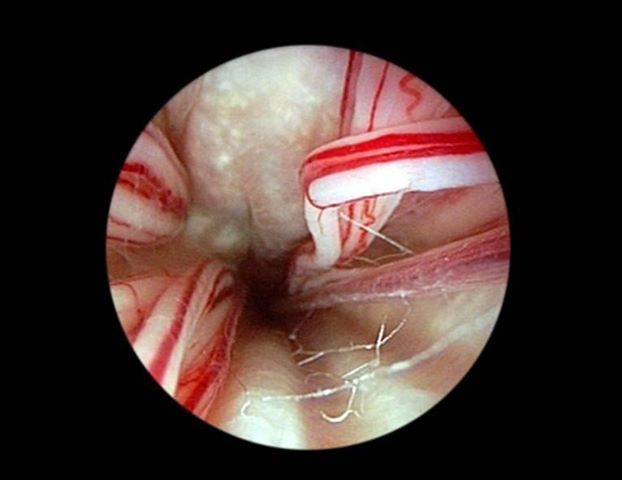 Picture taken of the inside of the spinal cord showing Adhesive Arachnoiditis |
 |
 |
 |
 |
 |
 |
 |
 |
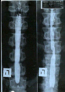
| MRI scan
showing Myodil clearly visible in the spinal
cord |
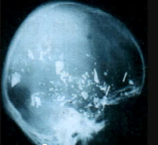
MRI scan of a victim, you can clearly see patches of myodil around the brain |
 |
 |
|
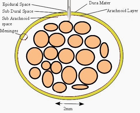
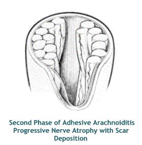
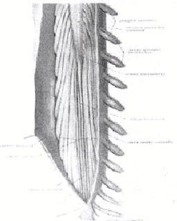
Diagram
showing
healthy spinal cord with nerve roots as they appear
naturally before chemical attack.
|
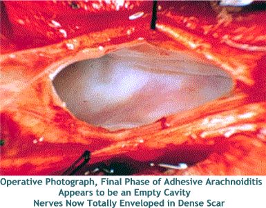
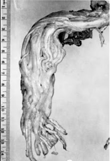
A spinal
cord taken from a victim, it has a candle wax appearance as
the nerves are encased in scar tissue. |
 |
 |
|
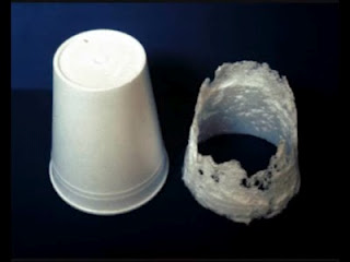
A plastic cup after
coming in to contact with myodil for just a
few hours.
|
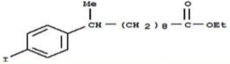
Myodil Chemical
Composition
Even
in
those early days it must have been a highly suspect
chemical mix considering the ingredients it used.
It consisted of Benzene (now regarded as the
number one carcinogenic in the world),
Hydrochloric Acid, Sulphuric Acid, Potassium
Permanganate (a substance that can kill if
digested) and 30% Iodine (if used incorrectly it can
lead to Parkinson's Disease).
|